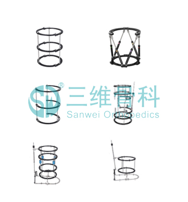
The Circular Fixator contains rings and rods of different sizes, which can be used in different positions. It is lightweight because of aluminium or carbon fibre construction. The enlongated hole give more flexibility and stability in nail putting.
Circular Fixator, External Fixator , Circular External Fixator, Ilizarov External Fixator
Primary useage: For Tibia & Femur Fixation
Certification:CE & ISO13485
Material:Aluminium
Advantages of our external fixators:
I. Unnular design, firm and reliable
II. Easier operation & short time
III. Minimally invasive surgery, no influence to blood supply of bone
IV. Second surgery is unnecessary, remove directly in clinic
V. Dynamic design, better for bone healing
VI. Taper bone screws, taut and firm after insertion.
We can also provide OEM service for you.
Hangwei Orthopedics Medical is a specialty medical device company that develops and markets products primarily for the Orthopedics.
We strive to provide superior benefits to professionals and patients through the development of reliable products.
We are a professional manufacturer of Circular Fixator/ Ilizarov Fixator, and look forward to cooperation with you!
Circular Fixator/ Ilizarov Fixator Circular Fixator,External Fixator,Circular External Fixator,Ilizarov External Fixator Shandong Hangwei Orthopedics Medcial Instrument Co., Ltd. , http://www.hangweimedical.com
1. The preparation of small pieces is the material basis of pearl formation. The quality of the small pieces is directly related to whether the pearl can be formed and its quality is good or bad. The extraction site and the shape, size, thickness and small piece production techniques of the tablets have a significant impact on the yield and quality of the bred pearls. The operation is as follows:
1.1 Cut open the shell with a knife to cut off the front and back of the small piece of muscle, shells open, wash mantle, visceral group and other dirt. Do not injure the outer membrane of the mantle and allow it to fit completely inside the clam shell.
1.2 Membrane separation The outer and inner epidermis of the limbic membrane were divided, and the outer epidermis of the limbic membrane was taken to prepare a small cell sheet. The surgical methods for separating the internal and external skins are stripping, tearing, and cutting.
1.2.1 The stripping method is to peel off the edge membrane on the clam shell to cut the outer skin. Use a pair of scissors to cut a small mouth in front of or behind the limbal membrane, clamp the inner surface of the epidermis with tweezers in one hand, and insert it into the inner and outer skin with a channel needle. All peeling of the epidermis. During the peeling process, the channel needle is as far as possible toward the side of the outer epidermis, so that the outer epidermis has less connective tissue, and a small uniform thickness is obtained. Then cut the outer epidermis with a slicing knife and place it on a glass slide with a forceps clip. Note that the pearl secretion surface is placed facing up.
1.2.2 tear membrane method is to first cut the edge of the membrane, the outer surface of the epidermis on the slide, press the end of the inner epidermis with a forceps, with a flat tweezers clip the same end of the outer skin, gently tearing along the tissue structure, Separate the outer and inner skins. Use a uniform force when tearing to prevent small tears or tears.
1.2.3 Cutting the membrane method Using a sharp scalpel, start from the front or back of the edge membrane, slowly cut off the epidermis and connective tissue of the mantle, and then cut the outer epidermis and place it on the slide.
Practice has proved that stripping membrane method is not difficult to operate, easy to grasp, but easy to damage the outer epidermal cells, the uneven thickness of the small pieces produced, affecting pearl yield and quality. Tear film method is currently the most commonly used method in production, technical difficulty is relatively large, small pieces of relatively uniform thickness, less damage, high quality, bred pearl brilliance, shape and yield are superior. Cutting film method is difficult to operate, slow speed, generally only applicable to pleated crowns, back corners no tooth tinea sheet. The performance of the three types of split membrane surgery and the formation of pearls (Yuan Zhu Wan is a spinnaker) are shown in Table 1.
Table 1 Comparison of the effects of different membrane separations and the performance of pearls. The rate of pearlization after transplantation. The pearl quality of pearls. The stripping method is easy to be slow and good. The circular tearing method is faster, faster, and better. The round cutting method is faster and difficult. smooth
1.3 Shaping and slicing Put the outer surface of the epidermis on the front (the pearl secretion surface on the shell side) facing up, and the opposite side (the surface of the connective tissue) facing down, spread flat on the glass plate, and trim it with a slicing knife to make it uniform in thickness and shape, especially This is the removal of the colored edge of the marginal membrane and the mantle muscle that remains on the inside of the marginal membrane. The size of the cut piece is generally 3mm4mm or 4mm5mm.
1.4 After the patch was made, a maintenance solution was dripped to keep the patch moist, maintain the balance of osmotic pressure inside and outside the cell, increase the viability and antibacterial function of the cell patch, and increase the survival rate of the cell patch.
Matters needing attention: The preparation of small pieces of action must be careful, light and skilled, do not hurt the outer epidermal cells; all tools before use with 75% alcohol cotton wipe disinfection; production process to prevent direct sunlight, so as not to affect the vitality of small pieces; keep the small pieces moist; The entire production process is completed in 3 to 5 minutes, keeping the vitality of the cells and avoiding the dry and small pieces caused by the long operation time.
2. After inserting small pieces to prepare small pieces, insert them into the connective tissue of the mantle of the cultured eubacteria, and heal them into one. Small pieces of planting steps: open the shell "fixed" cleaning mantle "pulling" to take tablets "wound" to send the film "whole circle" to change the side of the graft "plug" preparation.
2.1 Open the shell, fix the cultured bead with the belly upwards and place it on the operating frame. Use an opener to gently insert the beads between the two shells. Slowly open and insert the fixed plugs at both ends. Note that the width of the opening depends on the size of the carcass, and the length of the carcass is usually no more than 1.0 cm above 10 cm.
2.2 After washing the membrane and dialing clean water to wash the mantle and visceral sludge, use the duck tongue plate to move the iliac and visceral groups to the side that is not inserted.
2.3 insert (take tablets, wounds, to send tablets) There are two kinds of horizontal inserting method and straight inserting method.
2.3.1 Horizontal Insertion Method With the needle held in the left hand, gently hold the center of the small piece, and then hold the hook-shaped open needle in the right, place the small piece on the round head of the delivery needle so that the small piece is wrapped in a pouch shape, and then use the open crochet. Transverse wounds are made on the central membrane of the coat chamber, while the left hand provokes the small pieces to enter the connective tissue between the inner and outer epidermis of the mantle along the wound, and exits the crochet outside the inner epidermis to press the implanted piece to exit the delivery needle. The horizontal insertion method has the advantages of convenient operation, convenient rounding, high beading rate, good roundness, large number of inserts, and the like.
2.3.2 Vertical insertion method Vertical wounds are placed on the mantle membrane, and the small pieces are inserted perpendicular to the ventral edge of the cultured pearls. The in-line insertion method is easy to operate and can transplant larger pieces, but the number of inserts is small and it is easy to drop pieces. The number of grafts per lice depends on the size of the carcass. Over-consumption of the eucommia causes the eutrophic cervix to be overburdened and the nutrient supply is insufficient. Too little will affect the yield. General 6 ~ 8cm corpus callosum 2 ~ 3 rows, 10cm above the corpus callosum 4 rows, on both sides of the mantle can be implanted 30 ~ 40 pieces. The insert depth was 0.5-0.8cm, and the inter-patch distance was 0.5-1cm. The implanted small pieces were plum-type.
Remember that the delivery must be steady, light, and accurate. "One delivery" means that a small piece is implanted at a time to avoid multiple insertions and avoid wearing a membrane to prevent the appearance of enclosing shell beads.
2.4 Full circle When a small piece is implanted in the mantle, it is then pulled forward with a rounder or crochet on the outer surface of the outer surface of the epidermis, and the small pieces are formed into drum-like projections.
2.5 After the surgery on the mandibular membrane was changed on one side of the lateral implant, the direction of the ball was changed, and the zygomatic and visceral masses were dialed to the operated side with the duck tongue plate, and the other side was implanted.
2.6 Corking and preparation After completion of the operation, an antibacterial maintenance solution is added dropwise into the shell to prevent inflammation of the wound and promote healing. The plug was pulled out, and the cultured larvae were held in a microfluidic pool. After a period of observation, the larvae were collectively moved to a breeding pond for rearing.

Nucleation-free pearl cultivation surgical technique
Artificially cultivated pearls can be divided into seedless, nucleated, pictographic, atypical, colored pearls, and night pearls. The technique of fostering pearls is the key to artificial cultivation, and the level of technology is not relevant. It will seriously affect the yield and quality of the cultured pearls, and even result in the failure of the production of the cultured pearls. In order to prevent this kind of situation from happening and serve farmers better, this article details the operation methods and techniques for the cultivation of non-nuclear pearls.
